Basic HTML Version


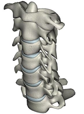
Cervical Spine
The Cervical spine is made up of 7 bones. You can
see on the MRI above on the right the bones sepa-
rated by the discs or shock absorbers. Behind the
bones you see a thick black cord, this is the spinal
cord. The Spinal cord comes down from the brain
and runs down into the spinal canal through the cer-
vical spine and thoracic spine where it ends.
There
is no spinal cord in the lumbar spine only nerves !
Thoracic Spine
The mid back or thoracic spine is made up of 12 ver-
tebral bodies which are connected to your 12 ribs.
Anywhere you feel a rib this is your thoracic spine,
above this is your Cervical spine and below this is
your Lumbar spine.
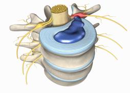
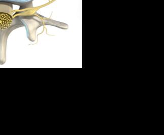
Nerves
At each level in the spinal nerves exit from the
spinal canal and send different types of signals to
our body. They provide signals of strength for our
muscles and transmit sensation from our skin. On
the photo above you can see how as the nerves
leave the spinal cord they have to travel alongside
the nearby discs. This is why disc herniations tend
to irritate our nerves.
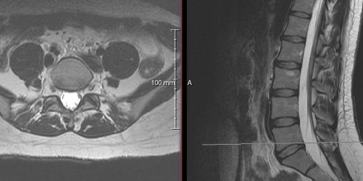
HELP ME UNDERSTANT THESE MRIs !!
This gets a little tricky and is best explained in person by
Dr. Pazmiño. On each MRI you will slices of the body
which appear as one of two different views: an Axial view
and a Sagittal view.
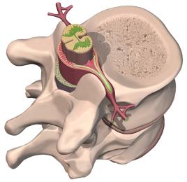
Axial View
This is the view on the left from the photo above. These
are horizontal images taken of your body as if you were
cut into slices by an axe. On these views you will see
your body in “cross section”. On the MRI above you will
see the nerves as white structures, compare this to the red
nerves displayed on the cartoon on the upper right. You
will also see a round white circle on the MRI which sits
behind the disc, this is seen on the cartoon photo as well
as the big round red cluster of nerves or spinal cord which
sits inside the brown bony spinal canal. As the nerves exit
the spine they encounter the disc in front and the joints of
the spine behind them. The
Facet Joints
are the joints of
the spine which control the motion of the spine similar to
how our knee joints control how much we can extend or
flex our knee. On the MRI these facet joints look like tiny
little hamburgers, do you see them? There are two of them
one on the left behind the nerve and one on the right side.
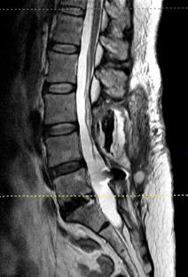
Sagittal View
The sagittal view is the side view of the spine. On the
sagittal view you should see the vertebral bones as blocks
which sit one atop the other. In between each block
you should see a white disc. MRIs are designed to show
objects with water as bright white substances. The discs of
your spine should be well hydrated and should appear on
the MRI as white shock absorbers set between the bony
blocks. Do you see these discs on the sagittal MRI images
above on the next page? Can you tell the difference
between healthy well hydrated white discs versus gray
/ black dehydrated and degenerated discs?
Once these
discs dehydrate they no longer function adequately
as shock absorbers which leads to back pain.
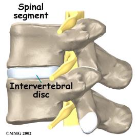
Discs
The discs are made up of two different materials.
The outer part of each disc is made up of a tough outer
ring of concentric fibers called the
Annulus Fibrosus.
These fibers serve to keep the shock absorber or the
Nucleus Pulposus
in place centrally.
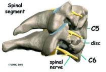
Here is one final photo demonstrating the relationship of
the nerves to the nearby discs in front and the facet joints
in the back.
FRONT
BACK
Degenerated Discs
Healthy Discs